42 heart structure and labels
Label the heart — Science Learning Hub In this interactive, you can label parts of the human heart. Drag and drop the text labels onto the boxes next to the heart diagram. If you want to redo an answer, click on the box and the answer will go back to the top so you can move it to another box. If you want to check your answers, use the Reset Incorrect button. Heart Labels - Printable or Custom Printed Stickers | Avery.com Use our free specialty shape label templates to easily personalize your heart labels online. Customize one of our free designs or upload your own graphics and then choose the printing option that works best for you. Order your blank heart labels or custom printed heart labels and stickers online and get free shipping on orders of $50 more.
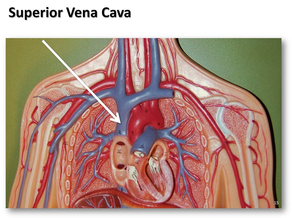
Structure of the Heart | The Franklin Institute The two largest veins that carry blood into the heart are the superior vena cava and the inferior vena cava. They are called "vena cava" because they are the "heart's veins." The superior is located near the top of the heart. The inferior is located beneath the superior. A wall called a septum, separates the right and left sides of the heart.

Heart structure and labels
Human Heart (Anatomy): Diagram, Function, Chambers, Location in Body The heart is a muscular organ about the size of a fist, located just behind and slightly left of the breastbone. The heart pumps blood through the network of arteries and veins called the ... quizlet.com › 630625176 › chapter-19-the-heart-flashChapter 19: The Heart Flashcards | Quizlet •Allows heart to beat without friction, gives it room to expand and resists excessive expansion •Parietal pericardium-tough outer, fibrous layer of connective tissue-inner serous layer •Visceral pericardium (a.k.a. epicardium of heart wall)-serous lining of sac turns inward at base of heart to cover the heart surface Label Heart Anatomy Diagram Printout - EnchantedLearning.com This cycle is then repeated. Every day, the heart pumps about 2,000 gallons (7,600 liters) of blood, beating about 100,000 times. Label the heart anatomy diagram below using the heart glossary. Note: On the diagram, the right side of the heart appears on the left side of the picture (and vice versa) because you are looking at the heart from the ...
Heart structure and labels. How to Draw the Internal Structure of the Heart (with Pictures) To finish drawing the aorta, draw three nubs at the top of the loop. After you draw these, erase the lines connecting from one side of the bottom of the nub to the other. Add tilted circles to the top of all of the nubs. Draw a circle at the bottom of the aorta, adjacent to the left ventricle. Heart Diagram with Labels and Detailed Explanation The heart is located under the ribcage, between the lungs and above the diaphragm. It weighs about 10.5 ounces and is cone shaped in structure. It consists of the following parts: Heart Detailed Diagram. Heart - Chambers There are four chambers of the heart. The upper two chambers are the auricles and the lower two are called ventricles. A Diagram of the Heart and Its Functioning Explained in Detail The heart blood flow diagram (flowchart) given below will help you to understand the pathway of blood through the heart.Initial five points denotes impure or deoxygenated blood and the last five points denotes pure or oxygenated blood. 1.Different Parts of the Body. ↓. 2.Major Veins. Structure of the Heart | SEER Training Structure of the Heart. The human heart is a four-chambered muscular organ, shaped and sized roughly like a man's closed fist with two-thirds of the mass to the left of midline. The heart is enclosed in a pericardial sac that is lined with the parietal layers of a serous membrane. The visceral layer of the serous membrane forms the epicardium.
The Anatomy of the Heart, Its Structures, and Functions The heart is the organ that helps supply blood and oxygen to all parts of the body. It is divided by a partition (or septum) into two halves. The halves are, in turn, divided into four chambers. The heart is situated within the chest cavity and surrounded by a fluid-filled sac called the pericardium. This amazing muscle produces electrical ... Heart Anatomy Labeling Game This is an online quiz called Heart Anatomy Labeling Game. There is a printable worksheet available for download here so you can take the quiz with pen and paper. Your Skills & Rank. Total Points. 0. Get started! Today's Rank--0. Today 's Points. One of us! Game Points. 19. You need to get 100% to score the 19 points available. Structure Of The Heart | A-Level Biology Revision Notes The heart is a hollow muscular organ that lies in the middle of the chest cavity. It is enclosed in the pericardium, which protects the heart and facilitates its pumping action. The heart is divided into four chambers: The two atria (auricles): these are the upper two chambers. They have thin walls which receive blood from veins. PDF Anatomy of Heart Labeled and Unlabeled Images (a) Anterior view of the external heart C' 2019 Pearson Education. Aort'c arch Ligamentum arteriosum Left pulmonary artery Left pulmonary ve ns Auricle of left atrium Circumflex artery Left coronary artery (in atrioventricular sulcus) Great cardiac vein Left ventricle Anterior interventricular artery (in anterior interventricular sulcus) Apex
Heart Labeling Quiz: How Much You Know About Heart Labeling? Here is a Heart labeling quiz for you. The human heart is a vital organ for every human. The more healthy your heart is, the longer the chances you have of surviving, so you better take care of it. Take the following quiz to know how much you know about your heart. 1. Human Heart - Diagram and Anatomy of the Heart - Innerbody Because the heart points to the left, about 2/3 of the heart's mass is found on the left side of the body and the other 1/3 is on the right. Anatomy of the Heart Pericardium. The heart sits within a fluid-filled cavity called the pericardial cavity. The walls and lining of the pericardial cavity are a special membrane known as the pericardium. byjus.com › biology › human-heartHuman Heart - Anatomy, Functions and Facts about Heart Following are the main functions of the heart: One of the primary functions of the human heart is to pump blood throughout the body. Blood delivers oxygen, hormones, glucose and other components to various parts of the body, including the human heart. The heart also ensures that adequate blood pressure is maintained in the body. Heart: Anatomy and Function Heart. Your heart is the main organ of your cardiovascular system, a network of blood vessels that pumps blood throughout your body. It also works with other body systems to control your heart rate and blood pressure. Your family history, personal health history and lifestyle all affect how well your heart works. Appointments 800.659.7822.
Pin by Daffodilcooper on BSC2086 | Heart model, Anatomy models labeled, Cardiac anatomy
Labelling the heart — Science Learning Hub Blood transports oxygen and nutrients to the body. It is also involved in the removal of metabolic wastes. In this interactive, you can label parts of the human heart. Drag and drop the text labels onto the boxes next to the diagram. Selecting or hovering over a box will highlight each area in the diagram.
Superior vena cava - The Anatomy of the Veins Visual Guide… | Flickr
› en › healthy-livingCarbohydrates | American Heart Association Carbohydrates are either called simple or complex, depending on the food’s chemical structure and how quickly the sugar is digested and absorbed. The type of carbohydrates that you eat makes a difference – Foods that contain high amounts of simple sugars, especially fructose raise triglyceride levels.

China U. S Trade War Heading To Economic Collapse : heading,News, breakingnews, globalnews ...
Diagrams, quizzes and worksheets of the heart | Kenhub Worksheet showing unlabelled heart diagrams. Using our unlabeled heart diagrams, you can challenge yourself to identify the individual parts of the heart as indicated by the arrows and fill-in-the-blank spaces. This exercise will help you to identify your weak spots, so you'll know which heart structures you need to spend more time studying ...
Drawing Of The Brain With Labels | Free download on ClipArtMag
Heart: illustrated anatomy - e-Anatomy - IMAIOS This interactive atlas of human heart anatomy is based on medical illustrations and cadaver photography. The user can show or hide the anatomical labels which provide a useful tool to create illustrations perfectly adapted for teaching. Anatomy of the heart: anatomical illustrations and structures, 3D model and photographs of dissection.

Histology Drawings: January 2014
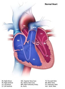
Heart Anatomy | Anatomy and Physiology - Lumen Learning Internal Structure of the Heart. Recall that the heart's contraction cycle follows a dual pattern of circulation—the pulmonary and systemic circuits—because of the pairs of chambers that pump blood into the circulation. In order to develop a more precise understanding of cardiac function, it is first necessary to explore the internal ...

Ask Thucydides! (“The Baker Street Irregulars’ ‘Thucydides’ whose Archival Series has set the ...
› heart-healthHeart Health | Heart Attack Prevention | Bayer® Aspirin TO HELP PREVENT ANOTHER HEART ATTACK. A doctor-directed aspirin regimen helps keep your blood flowing. Along with other heart-healthy choices, it can reduce your risk of having another heart attack. Learn About Aspirin's Benefits. Aspirin is not appropriate for everyone, so be sure to talk to your doctor before you begin an aspirin regimen.
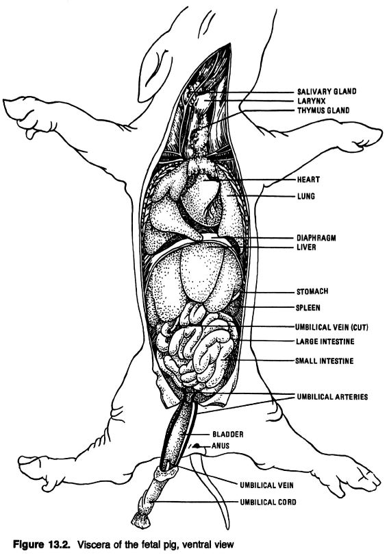
Anatomical Drawings of a Fetal Pig
variety.com › 2021 › musicOpen Letter Calls on Labels to 'Pay Songwriters' a More Fair ... Mar 23, 2021 · Andrew Lloyd Webber is among hundreds who signed an open letter calling on record labels to "Pay Songwriters" a more fair share. ... your artist would be a sensible and structure-preserving ...
How the Heart Works | Congenital Heart Defects | NCBDDD | CDC
Human Heart Diagram Labeled | Science Trends The pericardium is a structure that encases the heart, a sturdy structure made out of a double wall of tissue. The function of the pericardium is to hold the heart in a permanent place within the chest and give the heart protection. The outer layer of the pericardium is referred to as the parietal pericardium, and the serous pericardium is the inner layer of the structure.
Post a Comment for "42 heart structure and labels"