38 eye diagram with labels and functions
Human Eye: Structure of Human Eye (With Diagram) | Biology The human eye is a very sensitive and delicate organ suspended in the eye socket which protects it from injuries. It essentially consists of CORNEA, LENS & RETINA besides many other parts such as Iris, Pupil and aqueous humour, vituous humour etc. Each one has got a specific function. A section of the eye is as shown in Fig. 2.2. Human Eye Diagram, How The Eye Work -15 Amazing Facts of Eye First, light rays enter the eye through the cornea, the clear front "window" of the eye. The dome shaped cornea bends light to help the eye focus. From the cornea, the light passes through an opening called the pupil. The amount of light passing through is controlled by the iris, or the colored part of your eye.
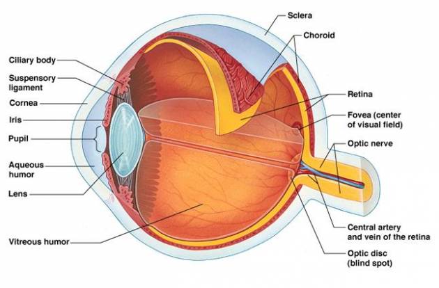
Structure and Function of the Human Eye - ThoughtCo The main parts of the human eye are the cornea, iris, pupil, aqueous humor, lens, vitreous humor, retina, and optic nerve. Light enters the eye by passing through the transparent cornea and aqueous humor. The iris controls the size of the pupil, which is the opening that allows light to enter the lens.

Eye diagram with labels and functions
The eye - Teaching resources Year 6 Light - Diagram of The Eye Labelled diagram by Ahamilton KS2 Y6 Science Structure and function of the Eye Open the box by Rworth Seasons wheel Random wheel by Kharris KS1 Science The Seasons Eye structures Balloon pop by Slloyd1 KS2 Biology Eye Labelling Labelled diagram by Swright Eye labeling Year 3 Labelled diagram by Sciencebowlingpark Microscope Types (with labeled diagrams) and Functions Simple microscope labeled diagram Simple microscope functions It is used in industrial applications like: Watchmakers to assemble watches Cloth industry to count the number of threads or fibers in a cloth Jewelers to examine the finer parts of jewelry Miniature artists to examine and build their work Also used to inspect finer details on products Eye Anatomy Diagram - EnchantedLearning.com Retina - light-sensitive tissue that lines the back of the eye. It contains millions of photoreceptors (rods and cones) that convert light rays into electrical impulses that are relayed to the brain via the optic nerve. Rods - cells the in the retina that sense brightness (they are photoreceptors). Night vision involves mostly rods (not cones).
Eye diagram with labels and functions. Eye muscles and their functions - All About Vision The main function of the extraocular eye muscles is to control eye movement and eye alignment. They are different from the intrinsic eye muscles, which enable the eye to focus on near objects and control how much light enters the eye. Extraocular eye muscles and their functions Structure and Functions of Human Eye with labelled Diagram Jun 24, 2021 · Cornea: It is the transparent, anterior or front part of our eye, which covers the pupil and the iris. The main function is to refract the light along with the lens. Iris: It is the pigmented, coloured portion of the eye, visible externally. The main function of the iris is to control the diameter of the pupil according to the light source. Labeled Eye Diagram | Science Trends The corneais the outermost portion of the eyeball. The responsibility of the cornea is to focus the light that enters our eyes. The cornea is transparent, and it covers the pupil, iris, and anterior chamber. The cornea itself is composed of five different layers, and the function of the outermost layer is to protect the eye from dirt and foreign ob... Eye Diagram With Labels and detailed description - BYJUS A brief description of the eye along with a well-labelled diagram is given below for reference. Well-Labelled Diagram of Eye The anterior chamber of the eye is the space between the cornea and the iris and is filled with a lubricating fluid, aqueous humour. The vascular layer of the eye, known as the choroid contains the connective tissue.
Generate eye diagram - MATLAB eyediagram - MathWorks 한국 Description. eyediagram (x,n) generates an eye diagram for signal x, plotting n samples in each trace. The labels on the horizontal axis of the diagram range between -1/2 and 1/2. The function assumes that the first value of the signal and every n th value thereafter, occur at integer times. eyediagram (x,n,period) sets the labels on the ... Eye Diagram Teaching Resources | Teachers Pay Teachers Students have to label a diagram of the eye. Students also have to identify the flow of light as it passes through the eye. They have to fill in the eye part and the function of the part. This activity supports Grade Seven California State Science Standard 5G - Students know how to relate the structures of the eye and ear to their functions. PDF Parts of the Eye - National Eye Institute | National Eye Institute To understand eye problems, it helps to know the different parts that make up the eye and the functions of these parts. Here are descriptions of some of the main parts of the eye: ... Handout illustrating parts of the eye Keywords: parts of the eye, eye diagram, vitreous gel, iris, cornea, pupil, lens, optic nerve, macula, retina ... Eye anatomy and function - AboutKidsHealth For people with normally functioning eyes, the following sequence takes place: Light reflects off the object we are looking at. Light rays enter the eye through the cornea at the front of the eye. The light passes through a watery fluid (aqueous humor), and enters the pupil to reach the lens.
Labelling the eye — Science Learning Hub In this interactive, you can label parts of the human eye. Use your mouse or finger to hover over a box to highlight the part to be named. Drag and drop the text labels onto the boxes next to the eye diagram If you want to redo an answer, click on the box and the answer will go back to the top so you can move it to another box. Microscope Parts and Functions With Labeled Diagram and Functions How ... Coarse adjustment: Brings the specimen into general focus. Fine adjustment: Fine tunes the focus and increases the detail of the specimen. Nosepiece: A rotating turret that houses the objective lenses. The viewer spins the nosepiece to select different objective lenses. Objective lenses: One of the most important parts of a compound microscope ... Cow's Eye Dissection - Eye diagram - Exploratorium A muscle that controls how much light enters the eye. It is suspended between the A cow's iris is brown. many colors, including brown, blue, green, and gray. A clear fluid that helps the cornea keep its rounded shape. The pupil is the dark circle in the center of your iris. It's a hole that Your pupil is round. Generate eye diagram - MATLAB eyediagram - MathWorks France Description. eyediagram (x,n) generates an eye diagram for signal x, plotting n samples in each trace. The labels on the horizontal axis of the diagram range between -1/2 and 1/2. The function assumes that the first value of the signal and every n th value thereafter, occur at integer times. eyediagram (x,n,period) sets the labels on the ...
Blank Eye Diagram - Cliparts.co
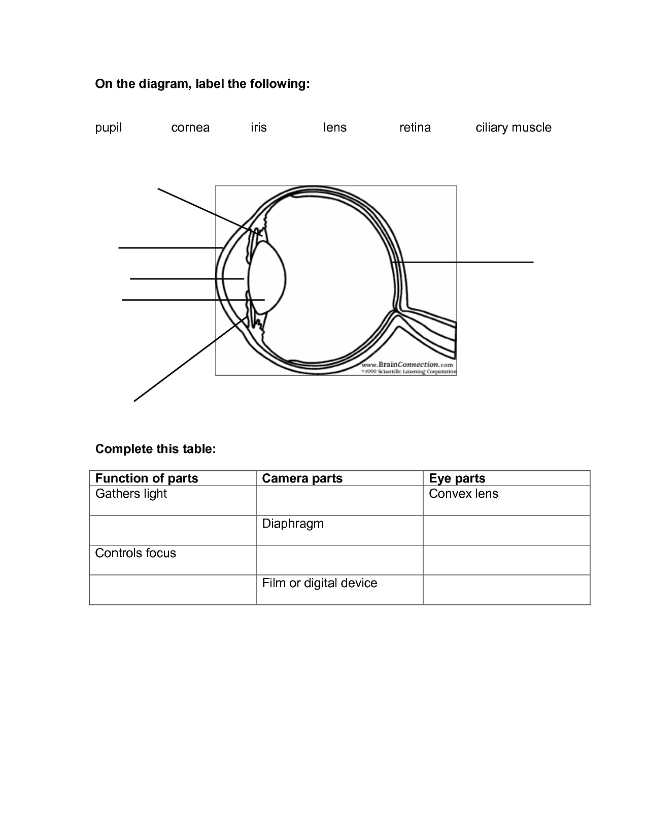
Label the Eye Worksheet - Teacher-Made Learning Resources - Twinkl In this resource, you'll find a 2-page PDF that is easy to download, print out, and use immediately with your class. The first page is a labelling exercise with two diagrams of the human eye. One is a view from the outside, and the other is a more detailed cross-section. On the second page, you'll find a set of answers showing the properly labelled human eyes, designed to help you check ...

GK Questions and Answers on Human Eye
Generate eye diagram - MATLAB eyediagram - MathWorks eyediagram (x,n,period) sets the labels on the horizontal axis to the range between - period /2 to period /2. eyediagram (x,n,period,offset) specifies the offset for the eye diagram. The function assumes that the ( offset + 1)th value of the signal and every n th value thereafter, occur at times that are integer multiples of period.
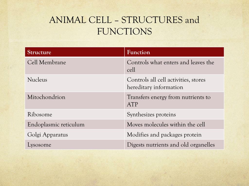
PPT - Cell Theory, Animal Cell Structure, Function, and Processes ...
Labelled Diagram of Human Eye, Explanation and Function - VEDANTU The basic functions of Rods and Cones are conscious light perception, color differentiation and depth perception. The human eye is capable of distinguishing between about 10 million colors, and it can also detect a single photo. The human eye is a part of the sensory nervous system. Labeled Diagram of Human Eye

Human Ear Diagram Blank - Diagram Media
Generate eye diagram - MATLAB eyediagram - MathWorks Italia eyediagram (x,n,period) sets the labels on the horizontal axis to the range between - period /2 to period /2. eyediagram (x,n,period,offset) specifies the offset for the eye diagram. The function assumes that the ( offset + 1)th value of the signal and every n th value thereafter, occur at times that are integer multiples of period.

parts – Graph Diagram
Parts of the Eye and Their Functions - Robertson Opt The different parts of the eye allow the body to take in light and perceive objects around us in the proper color, detail and depth. This allows people to make more informed decisions about their environment. If a portion of the eye becomes damaged, you may not be able to see effectively, or lose your vision all together.
8 Best Images of Lens Diagram Worksheet - Microscope with Labeled Parts ...
Human Eye Ball Anatomy & Physiology Diagram - eMedicineHealth Orbit. The orbit is the bony eye socket of the skull. The orbit is formed by the cheekbone, the forehead, the temple, and the side of the nose. The eye is cushioned within the orbit by pads of fat. In addition to the eyeball itself, the orbit contains the muscles that move the eye, blood vessels, and nerves.

8 Best Images of Lens Diagram Worksheet - Microscope with Labeled Parts ...
Eye anatomy: A closer look at the parts of the eye For more details about specific structures of the eye and how they function, visit these pages: Conjunctiva Of The Eye. Sclera: The White Of The Eye. Cornea Of The Eye. The Uvea Of The Eye. Pupil: Aperture Of The Eye. The Retina: Where Vision Begins. Macula Lutea Of The Eye. Choroid Of The Eye. Lens Of The Eye. Ciliary Body. Eye Muscles ...
Parts of the Eye and Their Functions | IYTmed.com
Diagram of the Eye - Home - Lions Eye Institute Instructions. Click the parts of the eye to see a description for each. Hover the diagram to zoom. Iris. The iris is the coloured part of the eye which surrounds the pupil. It controls light levels inside the eye, similar to the aperture on a camera. The iris contains tiny muscles that widen and narrow the pupil size.
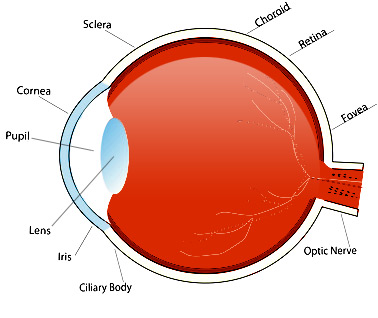
Diagram Of The Eye With Labels - ClipArt Best
PDF Eye Anatomy Handout - National Eye Institute of light entering the eye. Lens: The lens is a clear part of the eye behind the iris that helps to focus light, or an image, on the retina. Macula: The macula is the small, sensitive area of the retina that gives central vision. It is located in the center of the retina. Optic nerve: The optic nerve is the largest sensory nerve of the eye.
Post a Comment for "38 eye diagram with labels and functions"