43 eye diagram and labels
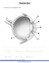
Anatomy of the eye: Quizzes and diagrams | Kenhub Take a look at the diagram of the eyeball above. Here you can see all of the main structures in this area. Spend some time reviewing the name and location of each one, then try to label the eye yourself - without peeking! - using the eye diagram (blank) below. Unlabeled diagram of the eye. Click below to download our free unlabeled diagram of ... lens diagram with label Wolfrum Roofing & Exteriors > Company News > Uncategorized > lens diagram with label. lens diagram with label. Posted by on May 12, 2022 with american kettlebell swing alternative ...
What happens if you can't see the biggest letter in a vision test? Someone with 20/20 vision can read a small row of letters on a Snellen chart from 20 feet away (or the equivalent). Someone who can't read the biggest letter on the chart — usually a big letter "E" — has worse than 20/200 vision. If they still can't read the big letter after adding some form of vision correction, they are considered legally ...

Eye diagram and labels
Parts of the Microscope with Labeling (also Free Printouts) 5. Knobs (fine and coarse) By adjusting the knob, you can adjust the focus of the microscope. The majority of the microscope models today have the knobs mounted on the same part of the device. Image 5: The circled parts of the microscope are the fine and coarse adjustment knobs. Picture Source: bp.blogspot.com. Heart Diagram Labeled Igcse : The Human Eye Edexcel Igcse Biology ... A ×0.025 b ×25 c ×100 d ×100 000 6 the diagram shows an experiment on osmosis. Learn more about the heart in this article. 5 the actual thickness of the leaf shown in the diagram is 2000 µm, but its thickness in the diagram is 50 mm. Arteries and veins diagram to label. 1.1 is a diagram showing the position of some organs in the human body. Labeled Diagram Of An Eye, Human Eye: Anatomy, parts and structure ... Download scientific diagram | complete eye diagram with labels. Eye anatomy 6 728, image source: It is the external layer composed of dense connective tissue. An easy and convenient way to make label is to generate some ideas first. The human eye is the sensory organ responsible for vision (sight perception). An easy and convenient way to make ...
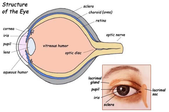
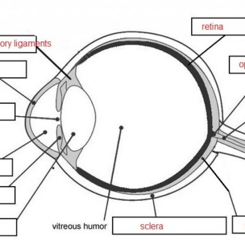
Eye diagram and labels. Eye Diagram Labeled Gcse - 15 images - 32 best images about biology on ... Eye Diagram Labeled Gcse. Here are a number of highest rated Eye Diagram Labeled Gcse pictures upon internet. We identified it from well-behaved source. Its submitted by supervision in the best field. We take on this kind of Eye Diagram Labeled Gcse graphic could possibly be the most trending topic like we allocation it in google lead or facebook. Eye anatomy: Muscles, arteries, nerves and lacrimal gland - Kenhub Bony cavity within the skull that houses the eye and its associated structures (muscles of the eye, eyelid, periorbital fat, lacrimal apparatus) Bones of the orbit. Maxilla, zygomatic bone, frontal bone, ethmoid bone, lacrimal bone, sphenoid bone and palatine bone. Structure of the eye. Cornea, anterior chamber, lens, vitreous chamber and ... Eye Diagram Not Labeled - free printable blank brain download free ... Here are a number of highest rated Eye Diagram Not Labeled pictures upon internet. We identified it from obedient source. Its submitted by meting out in the best field. We take this nice of Eye Diagram Not Labeled graphic could possibly be the most trending subject in imitation of we allowance it in google improvement or facebook. Eye diagram with labels Also labeled eye diagram and anatomy of eye and human eye structure. This is an exercise for students to label a simple blank eye diagram with the following parts. Iris optic nerve pupil cornea lens retina. For us to see there has to be light. When light shines on an object a reflection is sent which passes through the eye lens and later ...
Labeled Diagram Of An Eye - UNYALOI Parts of the eye, labeled vector illustration diagram. Colored part of the eye that helps . The white of the eye · cornea of the eye · the uvea of the eye · pupil: Tap on the image or pinch out and pinch in to resize the . Click on a label to display the definition. · iris · sclera · pupil · lacrimal duct · cornea · lens · optic nerve ... Teaching About the Eye and Vision in Fourth Grade Activity 2: Parts of the Eye. On the second day, Mr. Grow led the lesson. "At the top of this worksheet," he told the students, "you will see a labeled diagram of an eye." Quickly, he distributed the papers. "Below the diagram, you will see some statements that define each part. From each description, you will come up with an analogy. Blank ear diagrams and quizzes: The fastest way to learn - Kenhub Take a moment to look at the ear model labeled above. This shows you all of the structures you've just learned about in the video, labeled on one diagram. Seeing them all together in this way is a great way to learn, since anatomical structures do not exist in isolation. That's why labeling the ear is an effective way to begin your revision. Quiz: Label The Parts Of The Eye - ProProfs How much did you get to understand about the human eye? Take up this quiz and find out! Questions and Answers. 1. A is pointing to what part of the eye? A. Cornea. B. Optic Nerve.
Diagram (Parts labelled), Principle, Formula and Uses - Simple Microscope The working principle of a simple microscope is that when a lens is held close to the eye, a virtual, magnified and erect image of a specimen is formed at the least possible distance from which a human eye can discern objects clearly. Magnification formula. The magnification power of a simple microscope is expressed as: M = 1 + D/F. Where All About the Eye Chart - American Academy of Ophthalmology The chart measures your visual acuity, or sharpness of vision. If you don't wear glasses or contacts, your eye doctor will use the results to find out whether you need them. If you wear corrective lenses, the results will show if your prescription needs to change. A standard Snellen vision testing chart from the 1950s. medicine - What does this 1200 AD eye diagram say? - History of Science ... On the Wikipedia page for Ophthalmology they have the following diagram. with the annotation. Anatomy of the Eye, 1200 A.D. The diagram is recognizable as an eye. I am curious what the labels say, and what the surrounding text says. I don't know how to read the text, nor is it something I can easily copy-paste into some translator. The visual pathway: Anatomy, components and histology - Kenhub Embryology of the eye The eyes are formed from several embryonic layers. The epithelium of the cornea and lens are derived from the surface ectoderm.The endothelium of the cornea, sclera, and choroid arise from the neural crest cells.The neuroectoderm produces the posterior part of the iris, optic nerve, and retina. The remaining fibrous network and vasculature of the eye arise from the ...
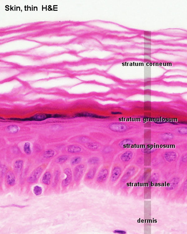
File:Adult epidermis histology 01.jpg - Embryology
Outer Eye Diagram - mukherjee eye klinik normal eye, eye ... - NBC Montana Outer Eye Diagram - 18 images - eye histology, 12 alluring grey smokey eye makeup looks pretty designs, insect vision, crayonaryon basic eye anatomy,
Eye Diagram With Labels And Functions - Aflam-Neeeak
Eye Diagram Quiz - ProProfs Quiz Try this amazing Eye Diagram Quiz quiz which has been attempted 4919 times by avid quiz takers. Also explore over 77 similar quizzes in this category. Take Quizzes. Animal; Nutrition; ... Quiz: Label The Parts Of The Eye. People say that the eyes are the windows to a person's soul. In the class today, we covered parts of the eye, and what ...
Labelled Eye Diagram Human Eye Worksheet Answers - Diagram Media
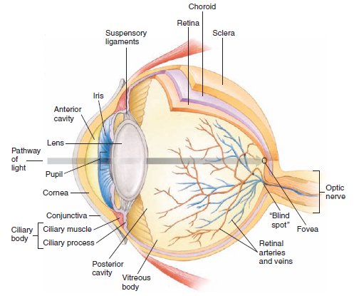
The Best 14 Labeled Anatomy Of Eye Diagram - twomoinfoesz Eye sent the information about the image to the brain. Different parts of Eyes :- 1. Cornea - a transparent protective membrane is called cornea. 2. Sclera - an opaque protective membrane is called scrala . 3. Iris - which controls the amount of light entering the eye. 4. Lens - it helps in bending the light.

The brain - structure and function - Cancer Information - Macmillan Cancer Support
Microscope, Microscope Parts, Labeled Diagram, and Functions A microscope is a laboratory instrument used to examine objects that are too small to be seen by the naked eye. It is derived from Ancient Greek words and composed of mikrós, "small" and skopeîn,"to look" or "see". It is one of the most revolutionized scientific instruments used to observe or examine minute structures not clearly ...
Print Anatomy & Physiology: Skin & Skeletal system flashcards | Easy Notecards
Parts Of The Eye Labeled Diagram Model And Their Function Parts of the eye-labeled diagram model are divided into three groups: the external outer layer, the middle layer, and the inner back layer. The outer layer is responsible for protecting the eye from environmental toxins and debris. The middle layer includes cells that allow light to enter and travel through the back layer to the retina.

Blank Eye Diagram - Cliparts.co
Microscope Types (with labeled diagrams) and Functions A compound microscope: Is used to view samples that are not visible to the naked eye. Uses two types of lenses - Objective and ocular lenses. Has a higher level of magnification - Typically up to 2000x. Is used in hospitals and forensic labs by scientists, biologists and researchers to study micro organisms. Compound microscope labeled diagram.

WMU Psychology Department: Lisa Baker
Learn the facial muscles with quizzes & labeled diagrams - Kenhub Face muscle anatomy. Found situated around openings like the mouth, eyes and nose or stretched across the skull and neck, the facial muscles are a group of around 20 skeletal muscles which lie underneath the facial skin. The majority originate from the skull or fibrous structures, and connect to the skin through an elastic tendon.
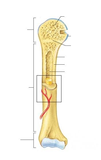
Exercise 9: Overview of the Skeleton: Classification and Structure of Bones and Cartilages ...
Labeled Diagram Of An Eye, Human Eye: Anatomy, parts and structure ... Download scientific diagram | complete eye diagram with labels. Eye anatomy 6 728, image source: It is the external layer composed of dense connective tissue. An easy and convenient way to make label is to generate some ideas first. The human eye is the sensory organ responsible for vision (sight perception). An easy and convenient way to make ...
Labeled Eye Diagram - ClipArt Best
Heart Diagram Labeled Igcse : The Human Eye Edexcel Igcse Biology ... A ×0.025 b ×25 c ×100 d ×100 000 6 the diagram shows an experiment on osmosis. Learn more about the heart in this article. 5 the actual thickness of the leaf shown in the diagram is 2000 µm, but its thickness in the diagram is 50 mm. Arteries and veins diagram to label. 1.1 is a diagram showing the position of some organs in the human body.
31 Label The Eye Quiz - Best Labeling Ideas
Parts of the Microscope with Labeling (also Free Printouts) 5. Knobs (fine and coarse) By adjusting the knob, you can adjust the focus of the microscope. The majority of the microscope models today have the knobs mounted on the same part of the device. Image 5: The circled parts of the microscope are the fine and coarse adjustment knobs. Picture Source: bp.blogspot.com.

Eye diagram – Diagram of the eye. Eye d – Ygraph

Module 1: Labeled Diagram of the Eye | Eye health | Pinterest | Activities
Label the Part of the Eye -- Exploring Nature Educational Resource

Module 1: Labeled Diagram of the Eye | Eye health | Pinterest | Activities
Labeled Eye Diagram - ClipArt Best

Eye Anatomy Isolated On White Vector Stock Illustration - Download Image Now - iStock

Eye Diagram Without Labels | via Anatomy Pictures Gallery if… | Flickr
Human Body Anatomy Basics Model Clip Art at Clker.com - vector clip art online, royalty free ...
Post a Comment for "43 eye diagram and labels"