45 brain mri with labels
Harvard University Show labels Show list All modalities to: MR-T1 MR-T2 FDG T1/FDG T2/FDG Perfusion MRI: The Five Most Frequently Asked Technical ... For DSC MR perfusion and DCE MR perfusion, we need to inject gadolinium-based contrast agent. The first MR contrast agent, gadopentetate dimeglumine (Magnevist, Bayer HealthCare), entered clinical trials for MRI of the brain in 1985 and was marketed initially in parts of Europe and Asia in 1988 and later in the United States. Since then, other ...
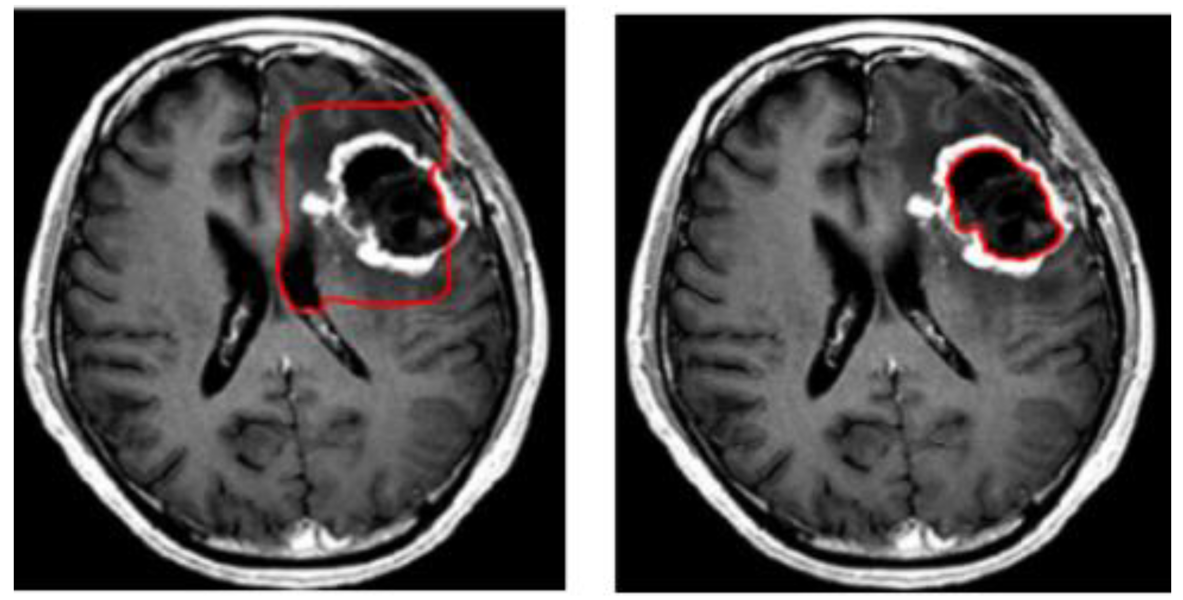
Brain Tumor Sequence Registration (BraTS-Reg) Challenge ... The data comprises of pairs of pre-operative baseline and follow-up MRI brain scans (each pair being of the same patient) diagnosed and treated for glioma. The exact multi-parametric MRI (mpMRI) sequences of each timepoint are i) native (T1) and ii) contrastenhancedT1-weighted (T1-CE), iii) T2-weighted and iv) T2Fluid Attenuated Inversion ...
Brain mri with labels
Cross-sectional anatomy of the brain - e-Anatomy - IMAIOS Apr 15, 2022 · Axial MRI Atlas of the Brain. Free online atlas with a comprehensive series of T1, contrast-enhanced T1, T2, T2*, FLAIR, Diffusion -weighted axial images from a normal humain brain. Scroll through the images with detailed labeling using our interactive interface. Perfect for clinicians, radiologists and residents reading brain MRI studies. precuneus: a review of its functional anatomy and behavioural ... Of particular interest are the neuroimaging studies seeking to define a physiological baseline state for the normal human brain function, since the precuneus shows one of the highest metabolic activity patterns of all brain regions during the conscious resting state and routinely exhibits decreases from this baseline across a variety of goal ... Researchers Automate Brain MRI Image Labeling Jul 28, 2021 ... Published in European Radiology, this is the first study allowing researchers to label complex MRI image datasets at scale. The researchers say ...
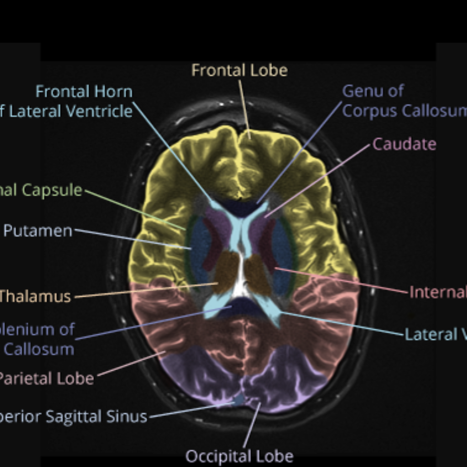
Brain mri with labels. Brain lobes - annotated MRI | Radiology Case | Radiopaedia.org Jul 14, 2018 ... Brain MRI with annotations of major structures. ADVERTISEMENT: Supporters see fewer/no ads. 13 articles feature ... Normal brain MRI - Kenhub MRI is used to analyze the anatomy of the brain and to identify some pathological conditions such as cerebrovascular incidents, demyelinating and ... MRI head axial T2 - labeling questions | Radiology Case Nov 2, 2021 ... The labeled structures are (excluding the correct side): cervical spinal cord posterior arch of C1 odontoid process (peg or dens) of C2 ... Researchers automate brain MRI image labelling, more than ... Jul 21, 2021 ... Researchers from the School of Biomedical Engineering & Imaging Sciences have automated brain MRI image labelling, needed to teach machine ...
MRI anatomy | free MRI axial brain anatomy This MRI brain cross sectional anatomy tool is absolutely free to use. This section of the website will explain large and minute details of axial brain ... Brain charts for the human lifespan | Nature Apr 06, 2022 · To extend the scope of brain charts beyond the four cerebrum tissue volumes, we generalized the same GAMLSS modelling approach to estimate normative trajectories for additional MRI phenotypes ... Automatic anatomical brain MRI segmentation combining label ... Oct 15, 2006 ... Regions in three-dimensional magnetic resonance (MR) brain images can be classified using protocols for manually segmenting and labeling ... The Whole Brain Atlas - Harvard Medical School Normal Brain: Normal Anatomy in 3-D with MRI/PET (Javascript) · Atlas of normal structure and blood flow ; Cerebrovascular Disease (stroke or "brain attack"):.
101 Labeled Brain Images and a Consistent Human Cortical ... Nov 14, 2012 ... An automated labeling system for subdividing the human cerebral cortex on MRI scans into gyral based regions of interest. Neuroimage 3, 968–980. Brain: Atlas of human anatomy with MRI - e-Anatomy - IMAIOS Sep 13, 2021 · MRI Atlas of the Brain. This page presents a comprehensive series of labeled axial, sagittal and coronal images from a normal human brain magnetic resonance imaging exam. This MRI brain cross-sectional anatomy tool serves as a reference atlas to guide radiologists and researchers in the accurate identification of the brain structures. Researchers Automate Brain MRI Image Labeling Jul 28, 2021 ... Published in European Radiology, this is the first study allowing researchers to label complex MRI image datasets at scale. The researchers say ... precuneus: a review of its functional anatomy and behavioural ... Of particular interest are the neuroimaging studies seeking to define a physiological baseline state for the normal human brain function, since the precuneus shows one of the highest metabolic activity patterns of all brain regions during the conscious resting state and routinely exhibits decreases from this baseline across a variety of goal ...
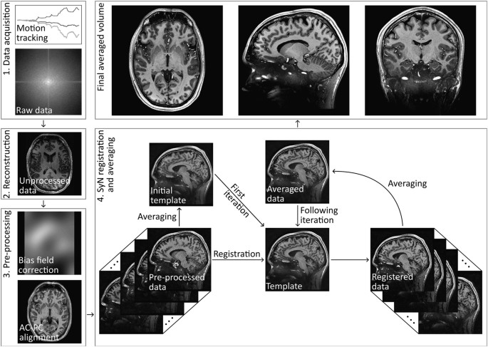
Design and fabrication of a realistic anthropomorphic ...
Cross-sectional anatomy of the brain - e-Anatomy - IMAIOS Apr 15, 2022 · Axial MRI Atlas of the Brain. Free online atlas with a comprehensive series of T1, contrast-enhanced T1, T2, T2*, FLAIR, Diffusion -weighted axial images from a normal humain brain. Scroll through the images with detailed labeling using our interactive interface. Perfect for clinicians, radiologists and residents reading brain MRI studies.
MRI identifies markers of atypical brain deve | EurekAlert!
T1-weighted in vivo human whole brain MRI dataset with an ...

Delaware Neuroscience - Brain Bee Detail, Page 2
Symmetry | Free Full-Text | 3D-MRI Brain Tumor Detection ...

Intelligent Scanning Using Deep Learning for MRI — The ...
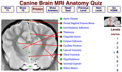
Veterinary Neurobiology Courseware
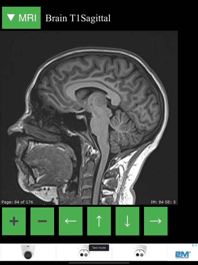
MRI Viewer on the App Store

Automatic brain labeling via multi-atlas guided fully ...

MRI anatomy | free MRI axial brain anatomy
CaseStacks.com - MRI Brain Anatomy

Brain Tumor Detection and Localization - Analytics Vidhya

MRI anatomy | free MRI axial brain anatomy

bio 151- mri human brain Diagram | Quizlet

volBrain: Automated MRI Brain volumetry system
Brain Imaging in Multiple Sclerosis: Practice Essentials ...

MRI anatomy | free MRI axial brain anatomy

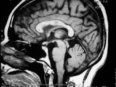
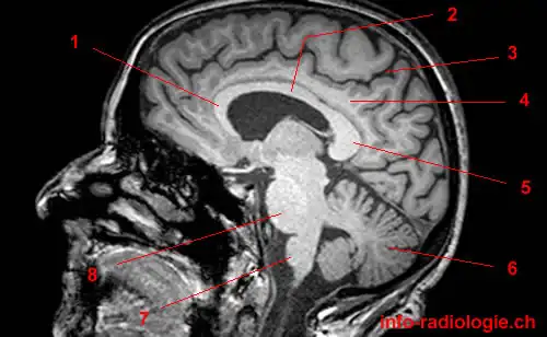
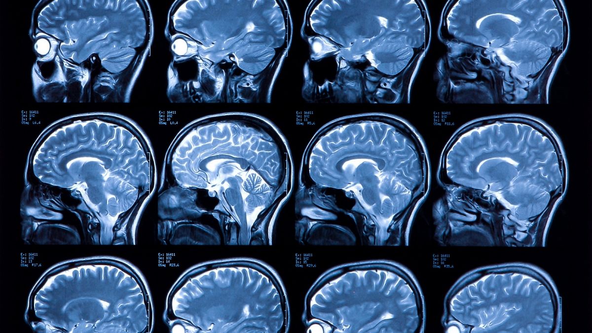
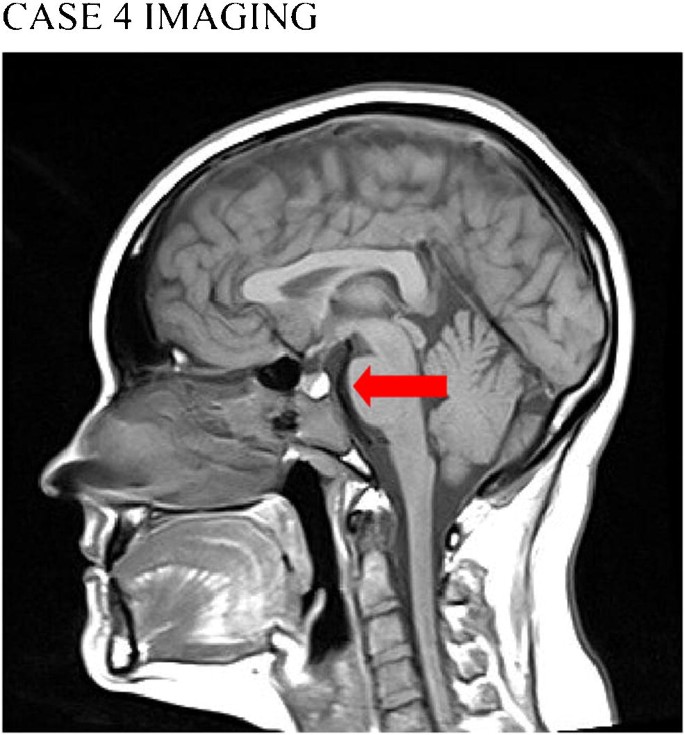
Normal anatomy of the brain on sagittal plane T1weighted ...
Atlas of BRAIN MRI - W-Radiology

Approach to MRI brain | LearningNeurology.com

Label Each Part of the Brain Scan | MS in African Americans ...

Cross-sectional anatomy of the brain - e-Anatomy
MRI scans prove useful for understanding depression

Tips and traps in brain MRI: Applications to vascular ...
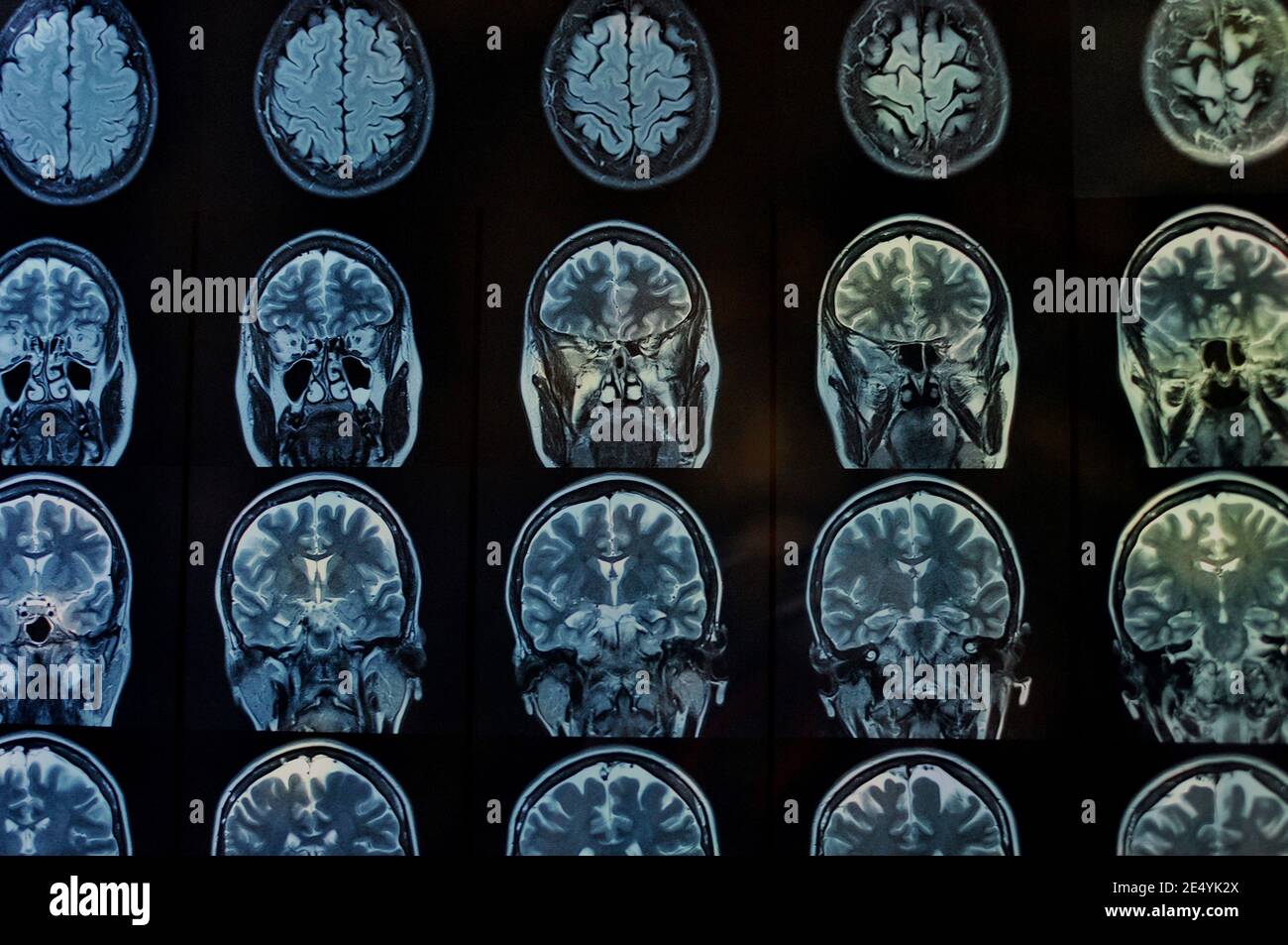
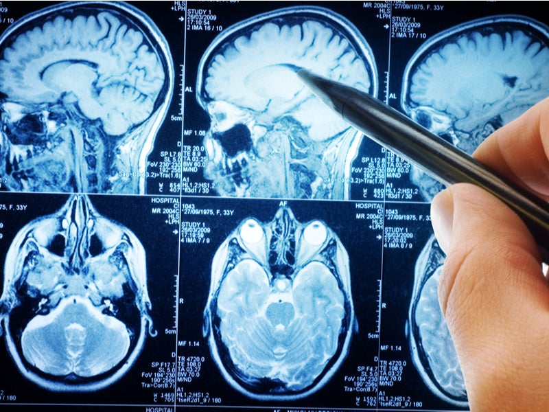
Brain scan hi-res stock photography and images - Alamy

Arterial Spin-Labeling Improves Detection of Intracranial ...
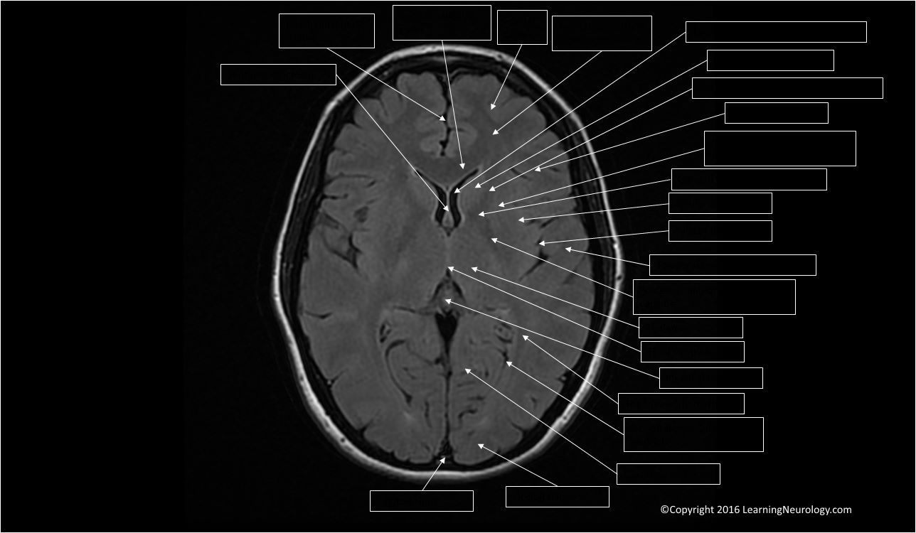
Approach to MRI brain | LearningNeurology.com

volBrain: Automated MRI Brain volumetry system

Cross-sectional anatomy of the brain - e-Anatomy

File:MRI brain sagittal section.jpg - Wikimedia Commons

Arterial Spin Labeling Perfusion of the Brain: Emerging ...

Read on for my tips at looking at a sagittal view MRI of the ...

MRI anatomy | free MRI axial brain anatomy

Labelled MRI of Normal Brain - Stock Image - C017/4418 ...
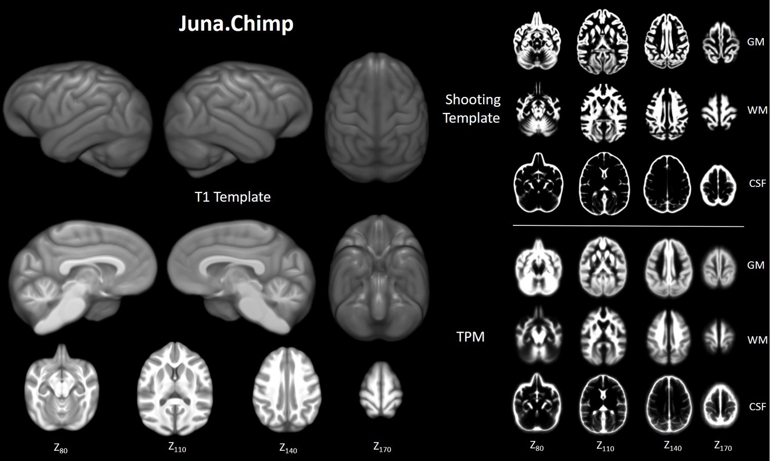
Chimpanzee brain morphometry utilizing standardized MRI ...

MRI anatomy | free MRI axial brain anatomy
Potentially life-saving study could cut labelling times for ...

How to Read a MRI of Brain - Brain Anatomy MRI Explained in English

Brain: Atlas of human anatomy with MRI - e-Anatomy
Brain: Atlas of human anatomy with MRI - e-Anatomy
Unusual Brain MRI Findings in Patients Imaged for Headache: a ...

MRI head axial T2 - labeling questions | Radiology Case ...

MRI Scans Show The Horrific Effect Cocaine Abuse Can Have On ...
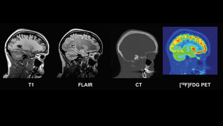
New database of healthy adult human brain PET, MRI and CT ...

MRI anatomy | free MRI axial brain anatomy
Post a Comment for "45 brain mri with labels"